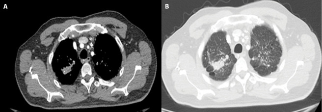
Figure 1.

42 years old man who has done sandblasting on glass for 20 years diagnosed as PMF.
A-B. Chest HRCT image shows irregularly shape with punctate calcification PMF lesion in the right upper lobe. Multiple small nodules indicative of silicosis are also seen in lung tissue bilaterally. Thick band appearance extending from the adjacent pleura to the PMF, and paracicatricial emphysematous lung tissue between the pleura and the PMF lesion was seen.
