
Fig. 8. DD Mtb can form without starvation, antibiotics, or limiting dilution.
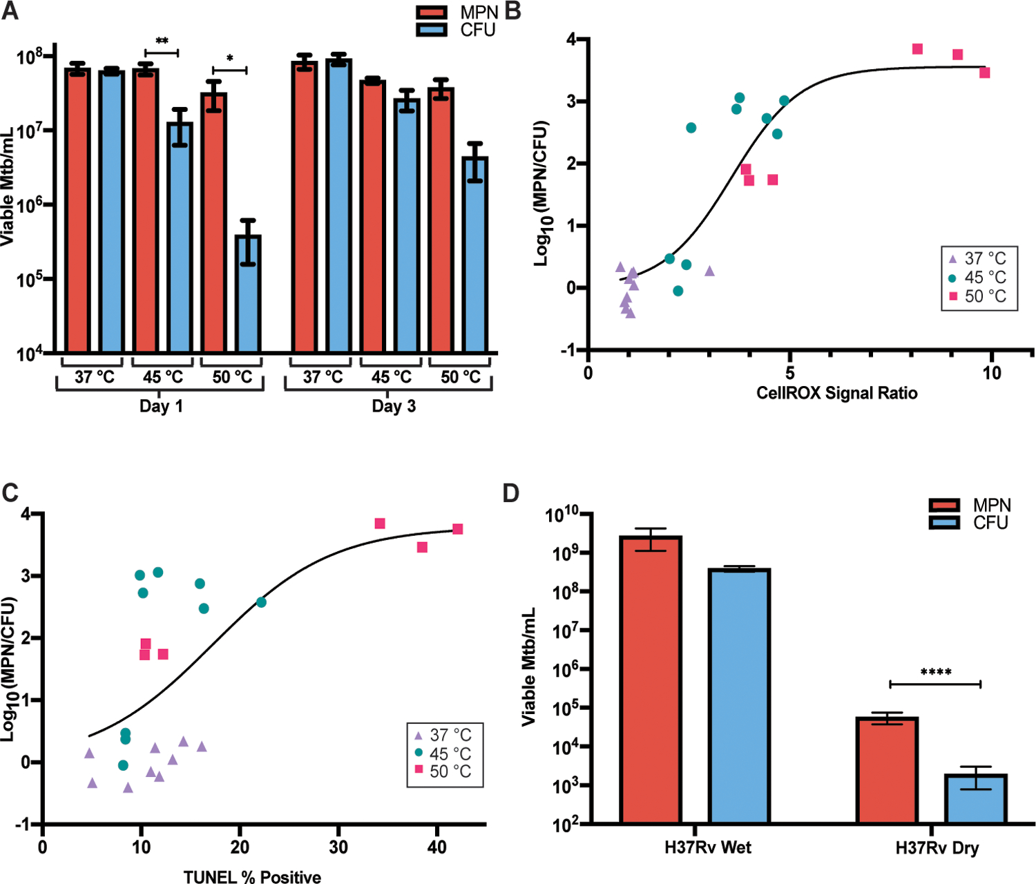
(A) Viable cell counts are shown for LP cells exposed for 24 hours to 37 °C, 45 °C and 50 °C immediately following heat exposure and after 48 hours at 37 °C. Data are representative of 3 experiments with 3 biological replicates each. Data were analyzed by a two-way ANOVA with Tukey’s multiple comparisons test. (B) CellROX % positive, normalized to average 37 °C CellROX % positive, is plotted versus proportion of DD Mtb after 1 day of indicated temperature exposures. Data are representative of 3 experiments with 3 biological replicates each and are shown with a nonlinear fit curve. (C) TUNEL positivity is plotted versus proportion of DD Mtb after 1 day of indicated temperature exposures. Data are representative of 3 experiments with 3 biological replicates each; one set of 50 °C was omitted due to extensive killing. Data are shown with a nonlinear fit curve. (D) Viable cell counts are shown for LP cells grown on filters and either desiccated or placed on a pool of saline for 7 days at 37 °C, 70% humidity, and room air CO2. Data are representative of 3 experiments with 2 to 3 biological replicates each. Data were analyzed by a two-way ANOVA with Sidak’s multiple comparisons test. All CFU were recorded and analyzed as the limit of detection, 100 CFU, if no growth was detected. *adj p < 0.05, **adj p < 0.01, ****adj p < 0.0001.