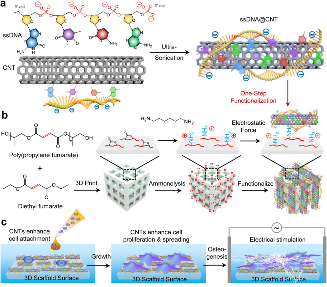
Fig. 1.
Schematic illustrations. a) Fabrication of ssDNA@CNT by ultra-sonication of ssDNA and CNTs. b) 3D scaffold–printing and subsequent ammonolysis and functionalization by ssDNA@CNT through electrostatic forces between positively charged 3D scaffold surfaces and negatively charged ssDNA@CNT composites. c) Enhanced cell proliferation and osteogenesis under electrical stimulation in the presence of ssDNA@CNTs.

