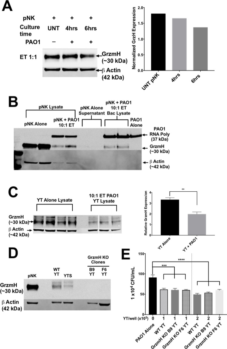
Fig 8. Single KO of GrzmH does not affect YT cell killing of PAO1.
(A) Western blot and densitometry of pNK cells (1x10⁶ cells), alone or co-cultured with 1x10⁶ cells bacteria for either 4 or 6 h. Blotted using (1:1000) mouse antibodies specific for human GrzmH. Rabbit antibody specific for human beta actin antibody was used as a loading control. Intracellular GrzmH levels were normalized to beta actin for densitometry. (B) Western blot analysis of pNK cells (1x10⁶ cells), alone or co-cultured with 1x10⁵ PAO1 for 6 h. PAO1 was isolated from the co-culture using a series of centrifugation and washes with sterile distilled water to form a bacterial pellet which was lysed with 2x Sample Buffer and then boiled. Blotted using (1:1000) mouse antibodies specific for human GrzmH. Rabbit antibody specific for human beta actin and a rabbit antibody specific was E. coli RNA polymerase were used as loading controls for the pNK and PAO1 cells respectively. (C) Western blot and densitometry of YT cells (5x10⁵ cells), alone or cultured with 4x10⁵ PAO1 for 6 h. Blotted using (1:1000) mouse antibodies specific for human Grzm H. Rabbit antibody specific for human beta actin antibody was used as a loading control. Intracellular GrzmH levels were normalized to beta actin for densitometry. (D) Western blot analysis of wild type or GrzmH KO YT cells (human leukemia NK cell line) (5x10⁵ cells) and blotted using (1:1000) mouse monoclonal antibodies specific for human GrzmH (4G5). Rabbit antibody specific for human beta actin antibody was used as a loading control. (E) PAO1 incubated alone or in the presence of wild type or GrzmH knockout YT cells and cultured for 6 h, then plated for CFU counts. Conditions were carried out in n = 4 wells (mean ± SEM) and the graph is representative of n ≥ 3 biological replicates * = P≤0.05, ** = P≤0.01, *** = P≤0.001.

