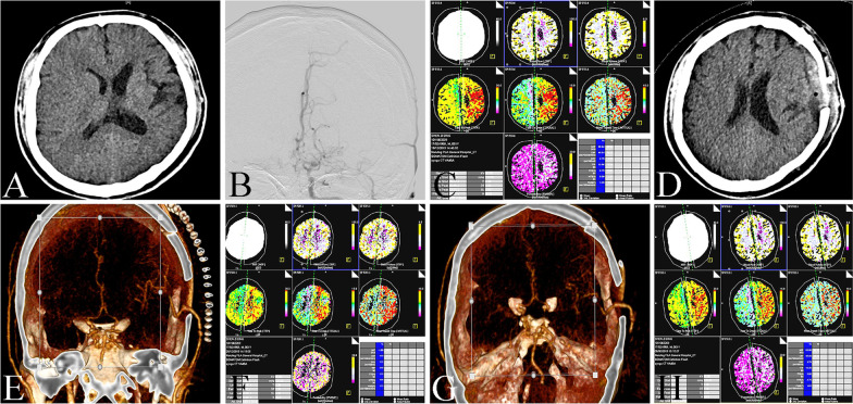
Fig. 2.
The image data of STA-MCA bypass. A Head CT showed infarcts in the left frontal lobe, corona radiata and centrum ovale. B DSA showed occlusion in left ICA. C CTP showed that the perfusion of left hemisphere was lower than that of right hemisphere. D Head CT showed that there was no new infarction or bleeding in the operation area. E CTA showed that the bypass vessels were unobstructed. F CTP showed that the left hemisphere perfusion was better than that before operation. G CTA showed that the bypass vessels were unobstructed after three months of follow-up. H CTP showed that left hemisphere perfusion was further improved after three months of follow-up

