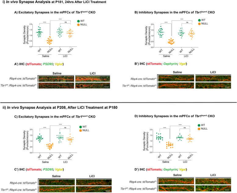
Fig. 1.
LiCl treatment restores normal synapse numbers in Tbr1layer5 mutant mice. Immunofluorescence (IF) was used to detect excitatory (A, C) and inhibitory (B, D) synapses onto dendrites from mPFCx of Tbr1wildtype (Rbp4-cre::tdTomatof/+; green), Tbr1layer5 CKOs (Tbr1f/f::Rbp4-cre::tdTomatof/+; orange) (n = 10 dendrites). Synapses were measured (i) 24 h and (ii) 4 weeks after injection with saline or LiCl at P180. Excitatory synapses were analyzed by VGlut1+ boutons and PSD95+ clusters co-localizing onto the dendrites from layer 5 neurons of mPFCx of Tbr1wildtype (green) and Tbr1layer5CKO (orange) mice 24 h (at P181; A, A’) and 4 weeks (P208; C, C’) after saline and/or LiCl was administered. Mann-Whitney ****p < 0.0001, ns = not significant. Inhibitory synaptic density was measured by VGat+ boutons and Gephyrin+ clusters co-localizing onto dendrites of mPFCx of Tbr1wildtype (green) and Tbr1layer5CKO (orange) 24 h (at P181; B, B’) and 4 weeks (P208; D, D’) after saline and/or LiCl was administered. Mann-Whitney ****p < 0.0001, ns = not significant. The Fiji ImageJ software was used to process confocal images for quantification. Two-tailed T-test with Mann-Whitney’s correction was used for pairwise comparisons

