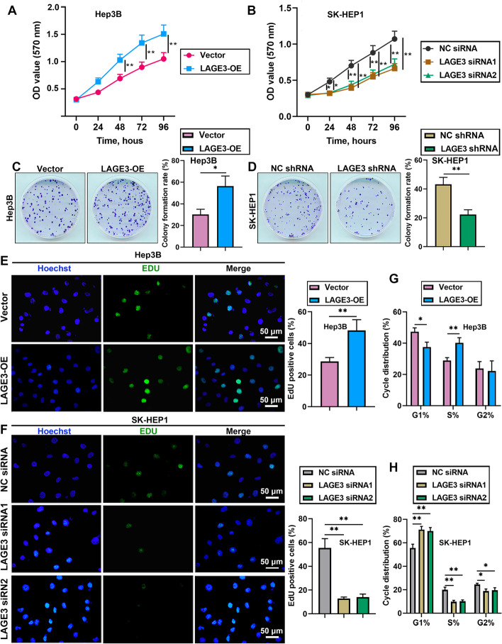
Fig. 2.
LAGE3 promoted the proliferation of HCC cells. Hep-3B cells were transfected with LAGE3-OE and SK-HEP1 cells were transfected with LAGE3 siRNA1/2 or LAGE3 shRNA. A–B MTT assay was used to evaluate the proliferation of Hep-3B and SK-HEP1 cells. C–D Colony formation assay was employed to assess the colony formation ability of Hep-3B and SK-HEP1 cells. E–F EdU-stained HCC cells. G–H Cell cycle analysis of HCC cells. Data are shown as mean ± standard deviation. *p < 0.05, **p < 0.01

