Fig. 6.
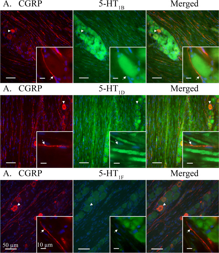
5-HT1B/D/F receptors co-staining with CGRP. CGRP-ir revealed a vesicular staining pattern in neuron cell bodies and C-fibres. A CGRP co-localized with 5-HT1B receptors in some neuron cell bodies (Arrowhead). No expression of the 5-HT1B receptor was observed in CGRP positive C-fibres. Insert: CGRP immunoreactive C-fibre adjacent to 5-HT1B positive neuron cell body. Arrow marks a visible C-fibre bouton. B CGRP-ir co-localized with 5-HT1D in neuron cell bodies (Arrowhead). No expression of the 5-HT1D receptor was detected in the CGRP immunoreactive C-fibres. Insert: CGRP positive C-fibre intermingled between 5-HT1D positive Aδ-fibres. Arrow marks a visible C-fibre bouton. C CGRP co-localized with weakly positive 5-HT1F receptors in neuron somas. No expression of the 5-HT1F receptor was detected in C-fibres. Insert: CGRP positive C-fibre in proximity to 5-HT1F positive neuron cell bodies. Arrow marks a visible C-fibre bouton

