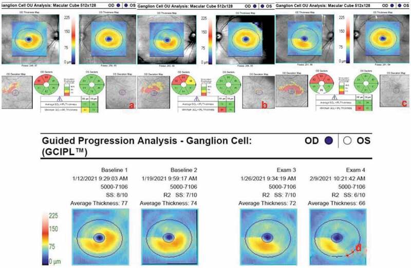
Figure 3.

Serial macular ganglion cell – inner plexiform layer (GCIPL) complex analyses of both eyes: (a) On day 55 ischaemic GCIPL loss of the supero-temporal fovea with a minimum thickness of 65 µm. The left GCIPL thickness is normal at 84 µm. (b) The same analysis repeated on day 62 reveals further atrophy enlarging supero-nasally. (c) Eventual atrophy involving the infero-nasal part of the macula can be seen on day 69. (d) Automated progression analysis showing ongoing GCIPL atrophy through 3 weeks until day 76.
