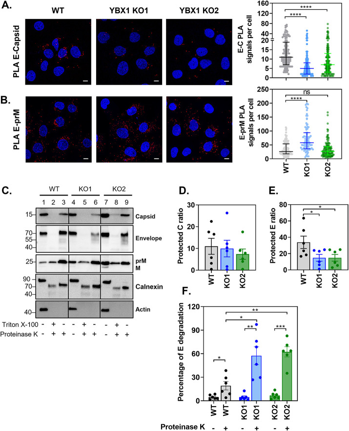
FIG 4.
YBX1 promotes the interaction between E and C proteins. (A–B) Huh7 WT and YBX1 KO cells were infected at MOI 5 and processed at 24 hpi for (A) E and capsid PLA and (B) and E and prM PLA. Nuclei were stained with DAPI (blue) and PLA signals are detected as red puncta. The number of PLA signals per cell were counted in at least 50 cells per experiment using an in-house macro for ImageJ. Data are presented as median ± IQR from two independent experiments. (C–F) Proteinase K (PK) protection assay of protein lysates from DENV-infected WT and YBX1-KO cells. Equal amounts of protein were left untreated or treated with PK in the absence and presence of Triton X-100 and samples were analyzed by Western blotting (C). The ratio of protected E protein (D) and C protein (E) was calculated by dividing the densitometry values of the PK-treated samples by that of the nontreated control. The percentage of E protein degradation (F) was calculated by dividing the densitometry value of the 70 kDa band by the densitometry value of the entire lane. Data are presented as mean ± SEM from six independent experiments.

