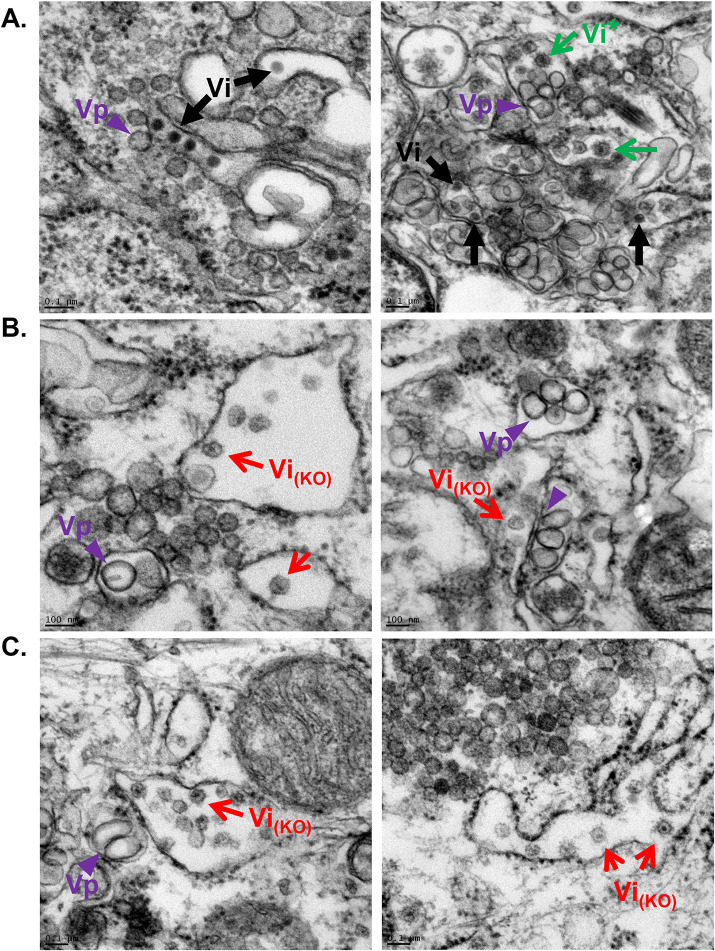
FIG 5.
Thin section TEM images of DENV-infected resin-embedded WT (A) and YBXI KO (B–C) Huh7 cells. Virus-induced structures include vesicle packets (Vp, purple arrowheads), which are the site of viral RNA replication. DENV infectious virions are identified as electron dense particles (Vi, black arrows) in arrays (A, left panel) and as individual virions (A, right panel). Empty looking particles with rough surface are detected in high abundance in the ER lumen of YBX1 KO cells (red arrows) and sporadically in WT cells (green arrows).

