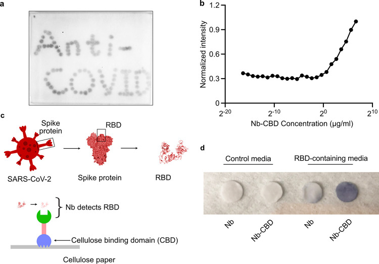
FIG 2.
The fusion protein maintains its activities in binding cellulose and the RBD of SARS-CoV-2. (a) Detection of immobilized Nb (Ty1)-CBD on a cellulose paper. Nb-CBD was first spotted onto a piece of cellulose paper. Upon air drying, the paper was incubated with a rat antibody against the FLAG epitope (DYKDDDDK), followed by an anti-rat secondary antibody conjugated with HRP. The dark precipitate “Anti-COVID” was visualized after incubation with 3,3′-diaminobenzidine (DAB). (b) Quantification of maximal protein absorption on Whatman filter paper. Ten microliters of serially diluted fusion protein solutions was applied to the filter paper, followed by immunoblotting with anti-FLAG directly on the filter paper. Based on the normalized unit intensity quantified by ImageJ, protein abundance increased with concentration. We estimate that 500 ng of Nb-CBD binds to 1 mm2 of cellulose paper at saturation status. (c) Schematic of an immunoassay to evaluate the function of the fusion protein. Nb-CBD fusion proteins were immobilized on cellulose paper and then submerged in culture medium containing RBD-Fc (∼100 ng/mL) as a proxy for actual SARS-CoV-2. The capture capability was confirmed by anti-human Fc-HRP and the DAB substrate. The structure of the RBD was adapted from PDB ID no. 6ZXN. (d) Testing the capture capability of protein-coated cellulose paper discs in RBD-containing medium. Representative discs were prepared by a 6-mm biopsy punch and then coated with E. coli lysates containing the indicated recombinant fusion proteins. The functionalized discs were incubated with RBD-containing or control (no RBD) medium. The intensity of dark staining was strongest from the combination of Nb-CBD-coated disc and RBD-containing medium (∼100 ng/mL).

