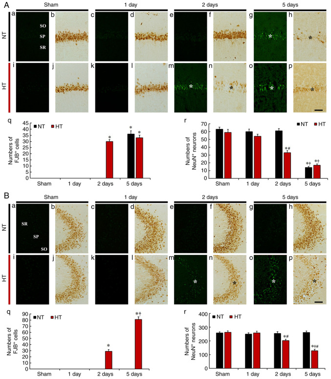
Figure 3.
(A and B) Fluoro-Jade B (FJB) histofluorescence staining and anti-neuronal nuclei (NeuN) immunohistochemistry in CA1 (A) and CA2/3 (B) of the NT/sham (a and b), NT/ischemia (c-h), HT/sham (i and j) and HT/ischemia (k-p) groups on day 1, 2 and 5 after TFI. In the NT/ischemia group, many FJB-stained cells (white asterisk in A-g) were observed in the SP on day 5 after TFI in CA1, but not in CA2/3. However, in the HT/ischemia group, many FJB-stained cells (white asterisks in m and o) were detected in both CA1 and CA2/3 from 2 days after TFI. The numbers of NeuN-immunostained pyramidal cells of the NT/ischemia group were significantly reduced (black asterisk in A-h) only in CA1 on day 5 after TFI. In the HT/ischemia group, the numbers of NeuN-immunostained pyramidal cells were decreased (black asterisks in n and p) in both CA1 and CA2/3 from 2 days after TFI. Scale bar, 50 µm. (q) Numbers of FJB-stained cells in CA1 (A) and CA2/3 (B). (r) Numbers of NeuN-immunostained cells in CA1 (A) and CA2/3 (B). *P<0.05 vs. NT/sham group; †P<0.05 vs. pre-time point group; #P<0.05 vs. NT/ischemia group. The bars indicate the means ± SEM (n=7, respectively). Note: the use of only small letters indicates these panels in both A and B.

