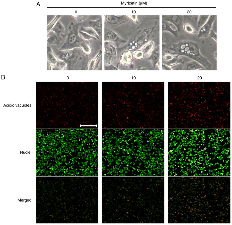
Figure 4.
Myricetin-induced morphological changes in SK-BR-3 cells. (A) Morphological changes (in particular, autophagic vacuoles) observed under a fluorescence microscope in SK-BR-3 cells treated with 10 and 20 µM of myricetin for 24 h. (B) Fluorescence microscopic images of SK-BR-3 cells stained with acridine orange to detect AVOs. The cytoplasm and the nucleus are stained fluorescent green, and the AVOs are stained fluorescent red. Scale bar, 10 µm. AVOs, acidic vesicular organelles.

