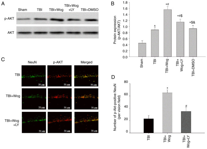
Figure 7.
Wogonin promotes the expression of p-Akt following TBI in the hippocampus through PI3K. (A) western blotting was performed on the levels of p-Akt and Akt in the hippocampal CA1 region of the sham group, TBI group, wogonin administration group (TBI + Wog), wogonin and LY294002 administration group (TBI + Wog + LY) and DMSO control group (TBI + DMSO) at 24 h after the operation. (B) The relative optical density of the p-Akt band was analyzed. (C) Representative immunofluorescence confocal images were shown to evaluate the colocalization of p-Akt (green) and NeuN (red). (D) The quantitative analysis of p-Akt-positive NeuN in the hippocampus. Scale bar, 50 µm. *P<0.05 vs. sham group; #P<0.05 vs. TBI group; $P<0.05 vs. TBI + Wog group; &P< vs. TBI + Wog + LY group. p-, phosphorylated; TBI, traumatic brain injury.

