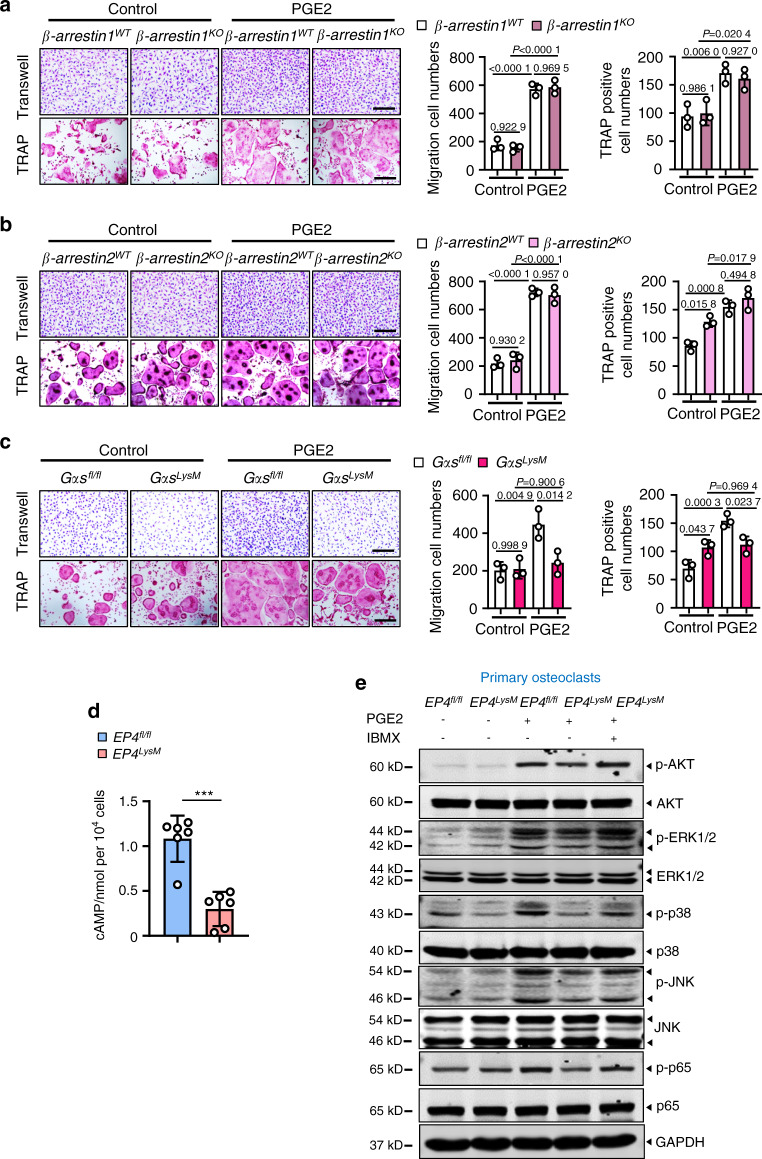
Fig. 8.
PGE2 regulates migration and osteoclast differentiation through the Gαs/PI3K/AKT/MAPK signaling pathways downstream of EP4. PGE2 regulates migration and osteoclast differentiation independent of β-arrestin1 (a) and β-arrestin2 (b) but via Gαs (c). BMMs from the WT and littermate β-arrestin1- and β-arrestin2-knockout mice were used to generate osteoclasts by stimulation with 10 ng·mL−1 M-CSF and 50 ng·mL−1 RANKL and incubation with 100 nmol·L−1 PGE2. For Gαs experiments, osteoclasts were generated from the GMMs of the Gαsfl/fl controls and Gαsfl/fl; LysM-cre (GαsLysM) mice. A differentiation assay (TRAP staining) (left) and the corresponding quantitative analysis (right). Scale bars, 50 μm. Error bars are the mean ± s.d. n = 3. Two-way ANOVA followed by Tukey’s t tests. Scale bars, 50 μm. d cAMP production was measured via ELISAs for osteoclasts derived from the Ep4fl/fl and Ep4LysM mice. BMMs from the two mouse strains were isolated and stimulated with 10 ng·mL−1 M-CSF and 50 ng·mL−1 RANKL to differentiate into osteoclasts and incubated with 100 nmol·L−1 PGE2 for 30 min prior to cAMP measurements. Error bars are the mean ± s.d. ***P < 0.001 by unpaired two-tailed Student’s t test. The experiment was performed with three biological replicates. e Representative images of the indicated protein expression by western blotting for osteoclasts generated using BMMs of Ep4fl/fl and Ep4LysM mice. The cells were treated either with osteoclastogenic media (10 ng·mL−1 M-CSF and 50 ng·mL−1 RANKL) alone, with PGE2 (100 nmol·L−1), or PGE2 with IBMX (1 mmol·L−1) for 3 h. The experiments were performed with three biological replicates

