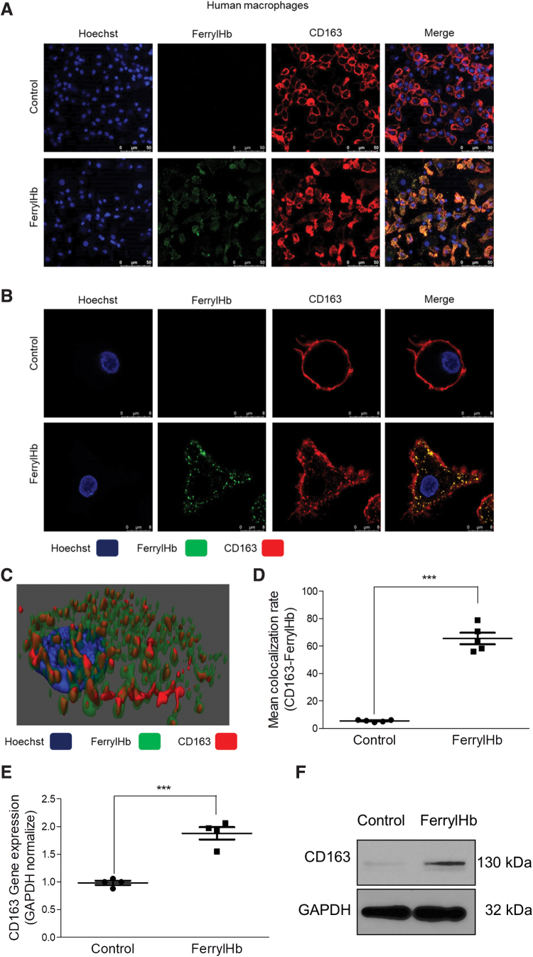
FIG. 11.
Ferryl hemoglobin and CD163 receptor are colocalized in macrophages exposed to ferryl hemoglobin. (A, B) Macrophages grown on coverslips were exposed to ferrylHb (10 μM) or growth medium for 16 h. Cells were stained with Hoechst 33258 for DNA (blue), an anti-ferrylHb antibody with Alexa Fluor 488 secondary antibody for ferrylHb (green), and an anti-CD163 antibody with Alexa Flour 647 secondary antibody for CD163 receptor (red). Images were taken with Leica TCS SP8 gated STED-CW nanoscopy. Images were deconvolved using Huygens Professional software. Representative image, n = 5. (C) 3D image demonstrated colocalization of ferrylHb and CD163 receptor. (D) The colocalization rate of ferrylHb and CD163 was calculated by ImageJ software (n = 5). (E) Macrophages were exposed to ferrylHb (10 μM) or growth medium for 16 h. Relative expression of CD163 in cells was analyzed by real-time qPCR (n = 5). (F) Macrophages were exposed to ferrylHb (10 μM) or growth medium for 16 h. The expression of CD163 was assessed by Western blot (n = 4). Scale bars shown in the images represent 25 or 50 μm. ***p < 0.001. Color images are available online.

