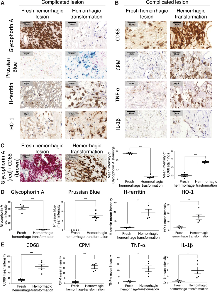
FIG. 2.
Cellular responses in human complicated plaque of the carotid artery. (A, B) The area of a fresh hemorrhagic region (A, B, left column) was compared with a hemorrhagic transformed region (A, B, right column) using immunohistochemistry. Glycophorin A positivity demonstrates intact red blood cells (fresh hemorrhage) within a lesion, whereas Prussian blue indicates iron accumulation (hemosiderin) in the hemorrhagic transformed area. The presence and activation of macrophages are indicated by markers of CD68, carboxypeptidase M/CPM, TNF-α, and IL-1β (at 100 × magnification). (C) CD68 and glycophorin A costaining was performed on complicated lesions. Images of fresh hemorrhagic area (left image) and hemorrhagic transformed region (right image) were shown. The mean intensity of CD68 and glycophorin A stainings was calculated using ImageJ software (N = 5). Heme-responsive proteins such as HO-1 and H-ferritin are demonstrated. (C–E) Quantitative analysis of immunohistochemical stainings of tissue sections was performed using ImageJ software (N = 5). Scale bars shown in the images represent 50 μm. **p < 0.01; ***p < 0.001. HO-1, heme oxygenase-1. Color images are available online.

