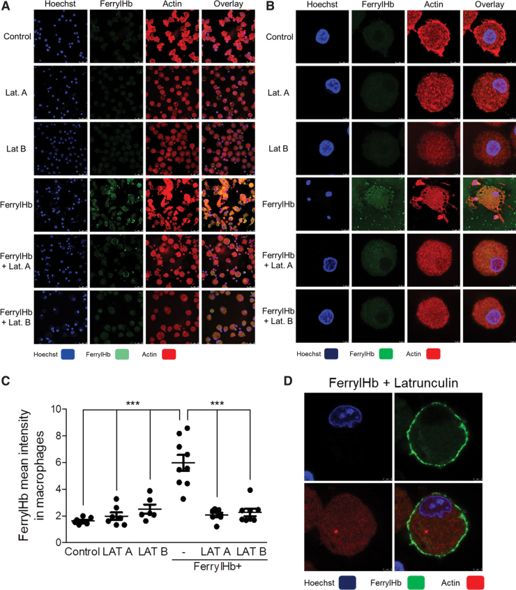
FIG. 8.
Inhibition of actin polymerization abrogates the uptake of ferryl hemoglobin by macrophages. (A, B, D) Macrophages were grown on coverslips and then treated with LAT A (500 nM); LAT B (10 μM) in the presence or absence of ferrylHb (10 μM); or growth medium for 16 h. Cells were stained with Hoechst 33258 for DNA (blue), an anti-ferrylHb antibody with Alexa Fluor 488 secondary antibody for ferrylHb (green), and Ifluor746 for actin (red). (A) Low-magnification and (B) high-magnification of images were shown. Images were taken with Leica TCS SP8 gated STED-CW nanoscopy. Images were deconvolved using Huygens Professional software. (C) FerrylHb intensity of macrophages was calculated by ImageJ software. Scale bars shown in the images represent (A, D) 3 μm and (B) 25 μm. ***p < 0.001. LAT A, latrunculin A; LAT B, latrunculin B. Color images are available online.

