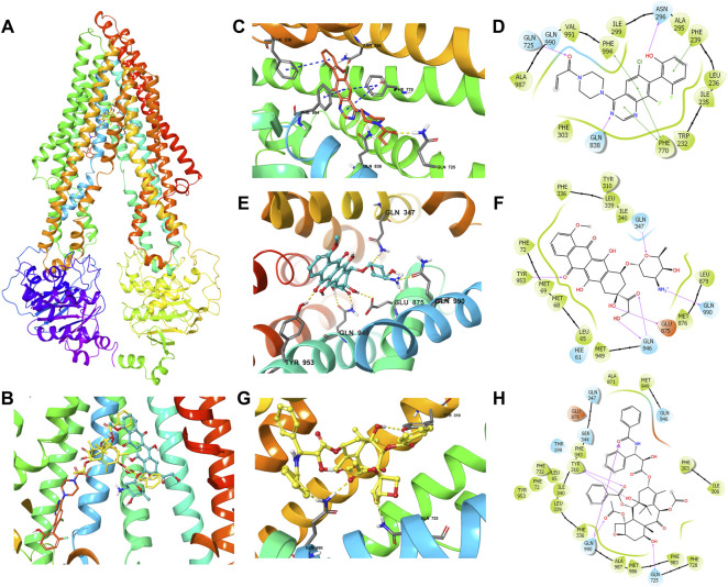
FIGURE 6.
The molecular modeling of ARS-1620, doxorubicin, and paclitaxel with human ABCB1. (A) The binding site of ARS-1620 (orange), doxorubicin (cyan), and paclitaxel (yellow) within human ABCB1. (B) The enlarged diagram of ARS-1620, paclitaxel, and doxorubicin within the binding site of human ABCB1. The interactions of ARS-1620 (C), doxorubicin (E), and paclitaxel (G) with the ABCB1 model in the three-dimension diagram. Ligands are shown as ball and stick mode: nitrogen—blue, oxygen—red, hydrogen—white, chloride—green, fluoride—light cyan. Interactions are indicated: hydrogen bonds—yellow dotted short line, π-π stacking—blue dotted short line. The interactions of ARS-1620 (D), doxorubicin (F), and paclitaxel (H) with the ABCB1 model in the two-dimension diagram: purple arrow hydrogen bond, green short line—π-π interaction.

