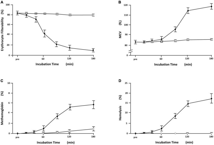
FIGURE 3.
Time-dependent derangement of erythrocytes exposed to 2,2′-azobis(2-amidinopropane) dihydrochloride (AAPH). Symbols and bars indicate mean ± SD (n = 6). (A) The filterability of erythrocytes exposed to 50 mM AAPH at 36°C showed time-dependent reduction ( ), which was marked at incubation time segment of 40–60 min after starting incubation, whereas the filterability of erythrocyte suspension without exposure to AAPH remained at the preincubation levels around 80% (◯). (B) Time-dependent changes of the mean corpuscular volume (MCV; fl) of erythrocytes with and without exposure to 50 mM AAPH. MCV is calculated automatically by hemocytometer as a surrogate of cell size. Erythrocytes exposed to 50 mM AAPH showed a sigmoidal increase in MCV (
 ), which was evident 60 min after starting incubation at 36°C. Erythrocytes without exposure to AAPH (◯) showed no discernible changes in MCV. (C) Time-dependent increases in methemoglobin formation of erythrocytes (%) with and without exposure to 50 mM AAPH. Methemoglobin was assayed by standard spectrophotometric methods using absorbance differences of hemolysate in the presence and the absence of potassium cyanide and/or potassium ferricyanide at the wavelength of 630 nm. Erythrocytes exposed to AAPH showed a time-dependent sigmoidal increase in methemoglobin formation (
 ), which was evident 60 min after starting incubation at 36°C. Erythrocytes without exposure to AAPH (◯) showed slight methemoglobin formation due to natural oxidation. (D) Hemolytic time course of erythrocyte suspension (%) exposed to 50 mM AAPH. Hemolysis was quantified by the absorbance of hemoglobin at 540 nm in the supernatant, and percent hemolysis (%) was calculated with comparison to complete hemolysis using distilled water. Erythrocytes exposed to AAPH (
 ) showed time-dependent hemolysis, which was evident 60 min after starting incubation at 36°C, whereas erythrocyte suspension without exposure to AAPH (◯) showed no evident hemolysis [cited from Odashiro et al. (2014) with permission].

