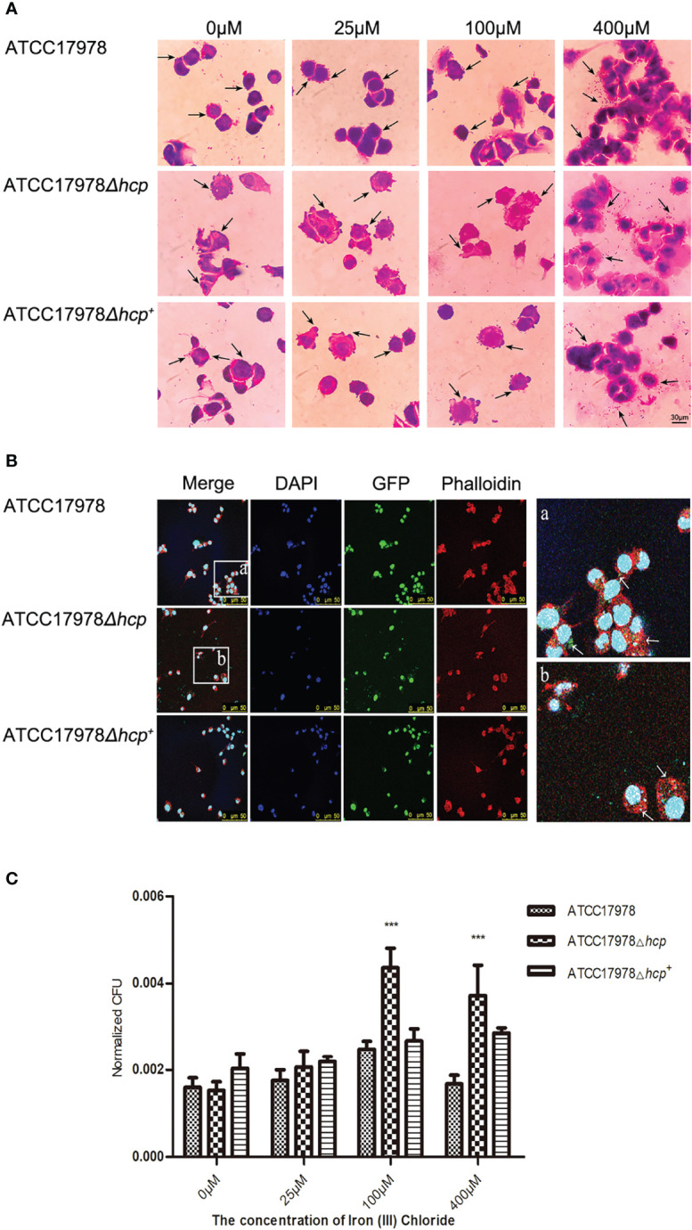
Figure 3.

Effect of iron on the Acinetobacter baumannii adhesion and invasion of human pulmonary alveolar epithelial cells (HPAEpiC). (A) Bacterial adhesion and invasion of the cells by the three strains (ATCC17978, ATCC17978Δhcp, and ATCC17978Δhcp+ ) in the presence of different concentrations of iron ions was observed under a conventional microscope. The arrows point to bacteria attached to the cell surface (not all marked, ×1,000). Scale bar = 30 μM. (B) Fluorescence microscopy of the effect of hcp on bacterial adhesion and invasion of the cells. The blue fluorescence represents cellular nucleic acids that were stained with DAPI; green fluorescent protein (GFP) was the fluorescent marker of bacteria, and the red fluorescence corresponds to a phalloidin staining of cellular actin, ×400. Schematic diagram of bacterial adhesion and invasion of HPAEpiC. The GFP labeling is indicated by arrows (not all marked). (C) The results of the bacteria count for bacteria adhesion and invasion of HPAEpiC by the three strains at different concentrations of ferric ion. *** represents means that are under the same iron ion. Compared with ATCC17978, ATCC17978Δhcp has a significant difference in normalized CFU adhesion and invasion of HPAEpiC (p < 0.001).
