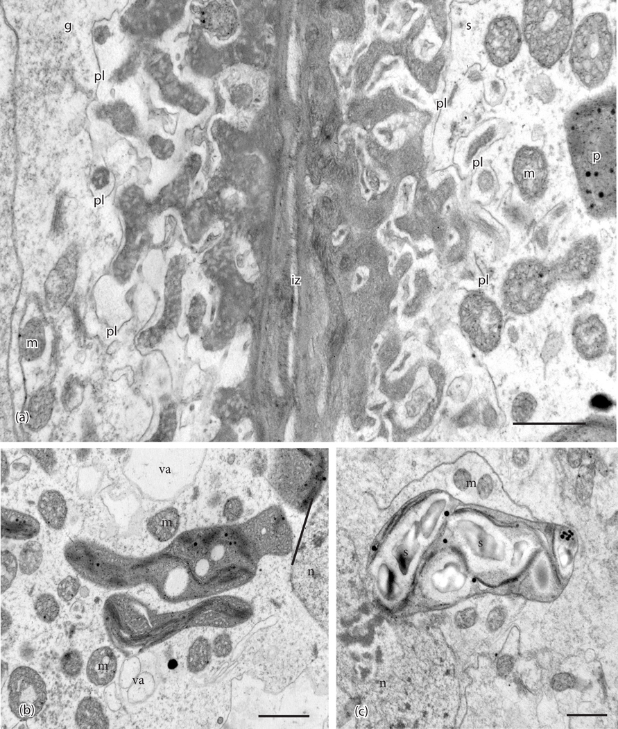
Figure 2.
Ultrastructural details of placental cells taken in the TEM. (a). Transfer cells in gametophyte (g) and sporophyte (s) are separated by a narrow intergenerational zone (iz) and show elaborate cell wall labyrinths. An electron-lucent region is delimited by the plasmalemma (pl) and surrounds the dense inner core of cell wall ingrowths that is more vesicular in the gametophyte. Mitochondria (m) and plastids (p) are located near wall ingrowths. (b). Plastids in sporophyte (s) cells are irregular in shape, dense, vesiculate, and contain few thylakoids. Mitochondria (m) and small vacuoles (va) are numerous in sporophyte cells. n, nucleus. (c). Plastids in gametophyte (g) cells contain starch grains (s) surrounded by thylakoids. m, mitochondria; n, nucleus. Scale bars = 0.1 μm

