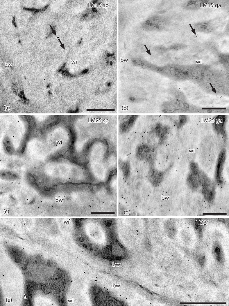
Figure 4.
TEMs of Physcomitrium patens placenta. Immunogold labeling with monoclonal antibodies to hemicellulose epitopes. (a). LM15 does not label the basal wall (bw) and sparsely labels (arrows) sporophyte cell wall ingrowths (wi). (b) LM15 does not label the basal wall (bw) and sparsely labels (arrows) gametophyte cell wall ingrowths (wi). (c). LM25 labels sporophyte placental cell wall ingrowths (wi) and the basal wall (bw). (d). LM25 labels gametophyte placental cell wall ingrowths (wi) and the basal wall (bw). (e). LM21 labels sporophyte (s) and gametophyte (g) transfer cell wall ingrowths (wi) and basal walls (bw). Scale bars = 0.5 μm.

