Figure 5.
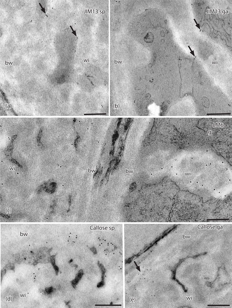
TEMs of Physcomitrium patens placenta. Immunogold labeling with monoclonal antibodies to AGP and callose epitopes. (a) In the sporophyte placental transfer cell, JIM13 labels (arrows) occur along the plasma membrane and wall ingrowths (wi) but not in the basal wall layer (bw). (b) Labels for JIM13 (arrows) occur in the gametophyte along the plasma membrane and wall ingrowths (wi) but not in the basal wall (bw). (c) LM6 labels are scattered throughout the wall ingrowths (wi) and basal wall (bw) in the sporophyte (s) and mostly in the electron lucent area along the edges of the wall ingrowths (wi) in the gametophyte (g) side, with few labels in the basal wall (bw). (d) Sporophyte and (e) Gametophyte. Labels for anti-callose (arrows) appear along the outer edge of the basal wall (bw) where it comes into contact with the wall ingrowths with few labels in wall ingrowths (wi). Scale bars = 0.5 μm.

