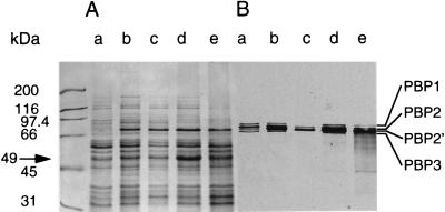
FIG. 5.
Membrane proteins and PBPs. (A) Coomassie blue-stained membrane proteins of strains BB255 (lane a), BB270 (lane b), BB270 (lane c), PG100 (lane d), and PG108 (lane e). All strains except strain BB270 in lane c were grown in LB medium without antibiotics. Strain BB270 in lane c was grown in the presence of 256 μg of methicillin ml−1. (B) Fluorography of the [3H]penicillin-labelled PBPs in the gel shown in panel A. The sizes of the molecular weight markers and the positions of the high-molecular-mass PBPs and of the 49-kDa band are indicated.

