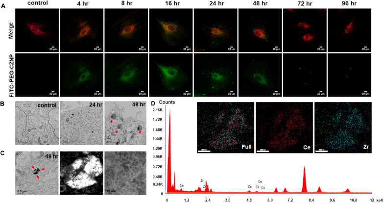
Fig. 2.
Intracellular localization and biodistribution of the PEG-CZNPs. A Confocal microscopy analysis of human podocyte treated with FITC-labeled PEG-CZNPs after 0, 4, 8, 16, 24, 48, 72 and 96 h. The human podocytes were then double-stained with Mitotracker (orange) and Lysotracker (blue). B TEM images of human podocytes treated with PEG-CZNPs after 0, 24, and 48 h. Residual PEG-CZNPs were found to be distributed in the lysosomes (red arrow). C STEM images of PEG-CZNPs in the lysosome of human podocytes D EDS spectra of PEG-CZNPs in the lysosomes of human podocytes to confirm their atomic compositions; 67 at% Ce and 32 at% Zr

