Fig. 4.
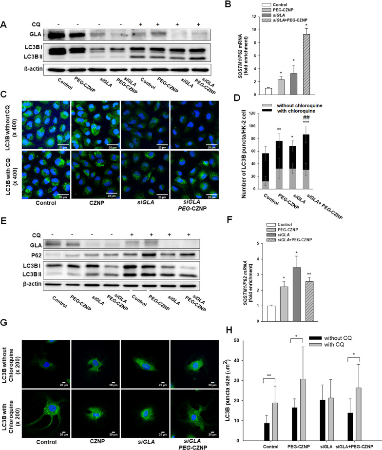
Effects of PEG-CZNPs on the autophagy response in HK-2 cells and in the human podocyte model of FD. A-D HK-2 cell analysis A Immunoblotting analysis of the autophagy response with and without chloroquine (CQ). B Quantitative real-time RT-PCR analysis of SQSTM1/P62 expression. C and D Confocal microscopy images of cells stained with LC3, with or without CQ exposure, and quantification of the fluorescence intensities using ImageJ software. E–H Human podocyte analysis E Immunoblotting analysis of the autophagy response with and without CQ exposure. F Quantitative real-time RT-PCR analysis of SQSTM1/P62 expression. G and H Confocal microscopy images of cells stained with LC3, with or without CQ exposure, and quantification of the fluorescence intensities using ImageJ software. Data values represent the mean ± SD; *P < 0.05, **P < 0.01, and ***P < 0.001 versus control; ###P < 0.001 versus siGLA knockdown alone

