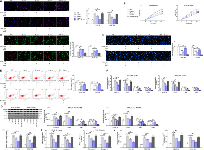
Fig. 2.
ucMSC-Evs suppress PDGF-induced proliferation of rMCs. A proliferation of rMCs determined by EdU labeling; B viability of cells determined using a CTG kit; C portion of dead cells confirmed by EB fluorescence staining; D apoptosis of cells determined by Hoechst 33,258 staining; E, apoptosis rate of cells determined by flow cytometry; F, G mRNA (F) and protein (G) expression of Cyclin D2, PCNA, Bax and PUMA in cells determined by RT-qPCR and western blot analysis, respectively; H, total collagen concentration in rMCs determined by collagen kits; I, J, expression of fibronectin-1 and collagen IV in cells determined by RT-qPCR and immunofluorescence staining. Data were expressed as mean ± SD from at least three independent experiments. Data were analyzed by two-way ANOVA followed by Tukey’s multiple comparison test. **p < 0.01, ***p < 0.001 versus PBS group; ##p < 0.01, ###p < 0.001 versus 0 μg/mL group

