. 2022 Jan 11;13(10):2857–2876. doi: 10.1039/d1sc06269g

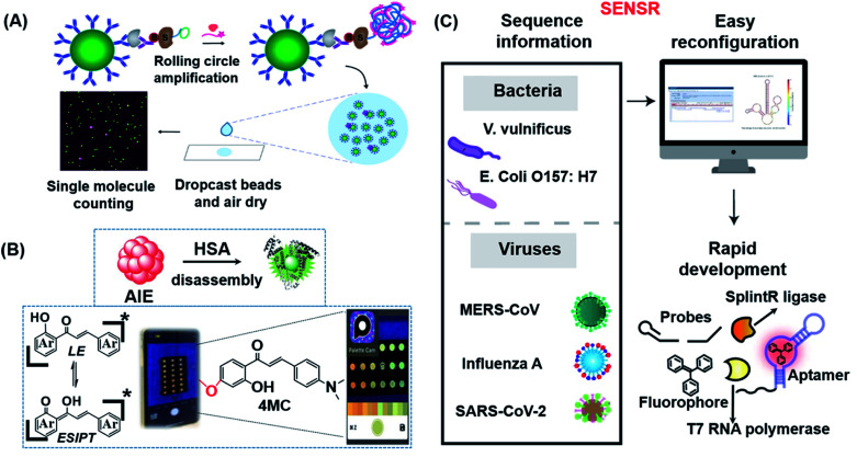
Fig. 9. (A) Schematic illustration of dSimoa. From left to right shown in the scheme, the main structure was composed of antibody-coated paramagnetic beads, the target protein, the biotinylated detector antibody, the labeled streptavidin–DNA conjugate, DNA polymerase and the probe. RCA is conducted on the beads after the development of a single immunocomplex on antibody–magnetic beads–streptavidin–DNA sandwiches to construct a lengthy concatemer connected to each immunocomplex. The concentrated beads are then dropcast onto a microscope slide and fluorescence imaging of the dropcast film is used to count single target molecules. Reproduced from ref. 149 with permission. Copyright 2020 American Chemical Society. (B) Schematic showing the design of the paper-based ratiometric fluorescence analytical device and the chemical structure of 4MC. In this assay, the concentration of HSA was determined by comparing the ratios of emission from probes in aggregated and monomeric states, which results in a visible red-to-green color change on a simple, portable paper-based analytical device developed by integrating the identification probe into the detecting pad. Reproduced from ref. 150 with permission. Copyright 2020 Wiley-VCH. (C) Illustration of the broad adaptability of SENSR and its easy reconfiguration and rapid development for various pathogens. The assay relies on a sustained isothermal reaction cascade producing an RNA aptamer that binds to a fluorogenic dye. T7 RNA polymerase transcribes the RNA aptamer from a promoter DNA probe and a reporter DNA probe that hybridize with the target single-stranded RNA sequence via the SplintR ligase. After the one-pot isothermal reaction, the complex shows strong fluorescence in the reaction tube. Two pathogenic microbes and three viruses were targeted by redesigning the probe sequences. Reproduced from ref. 151 with permission. Copyright 2020 Springer Nature.

Fig. 9