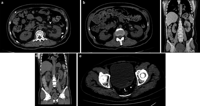
Figure 7.
a: NCCT KUB (axial section) depicting an enlarged and bulky left kidney (arrow) with surrounding peri-nephric fat stranding (arrowhead). b: NCCT KUB (axial section) depicting an enlarged left ureter (arrowhead). c: NCCT KUB (coronal section) depicting an enlarged and bulky left kidney (arrowhead) with surrounding perinephric fat stranding. d: NCCT KUB (coronal section, maximum intensity projection image) depicting a left-sided DJ stent (arrowhead). e: NCCT KUB (axial section) depicting bladder wall thickening (arrow) with prominent and hyperdense appearing left VUJ (arrowhead). NCCT KUB, non-contrast CT of Kidneys, ureters, and bladder

