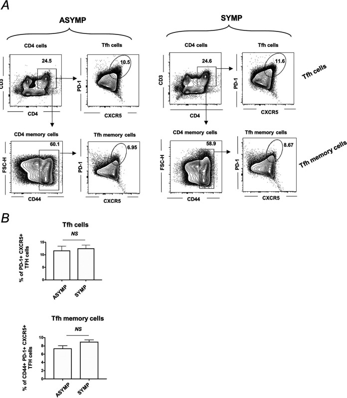
FIG 6.
T follicular helper (Tfh) and T follicular helper memory (Tfh memory) cell profile in spleen of ASYMP and SYMP HSV-1-infected mice. B6 mice (n = 6) were infected with HSV-1 McKrae (1 × 10⁶ PFU/eye) by scarification, and at day 35 p.i., reactivation was done by 60 s corneal UV-B irradiation. Mice were categorized as ASYMP/SYMP and euthanized at day 6 postreactivation. Spleen cells were collected for flow cytometry staining of Tfh (CD3+CD4+CXCR5+PD-1+ cells) and Tfh memory (CD3+CD4+CD44+CXCR5+PD-1+ cells). (A) Representative FACS plots for Tfh cells (CD3+CD4+CXCR5+PD-1+ cells) are shown in the top panel, and Tfh memory cells (CD3+CD4+CD44+CXCR5+PD-1+ cells) are shown in bottom panels for ASYMP (Left) and SYMP (Right) infected mice. (A) Graph showing percentage of Tfh cells (top) and Tfh memory cells (bottom) in spleen of ASYMP and SYMP infected mice is shown.

