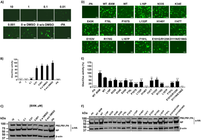
FIG 7.
Effect of BXM in A/Viet Nam/1203/2004 H5N1 PA polymerase activity. (A to C) Effect of BXM in polymerase activity of replication complexes containing the WT PA. Human HEK293T cells were transiently cotransfected (96-well plate format, 1 × 10⁴ cells/well, triplicates) with 31.25 ng of A/Viet Nam/1203/2004 H5N1 pCAGGS expression plasmids encoding HA-tagged PB2, PB1, PA, and NP, together with 62.5 ng of vRNA-like Gluc or GFP expression plasmids (pPOL-I) under the control of the human polymerase I promoter; and 12.5 ng of the SV40-Cluc plasmid to normalize transfection efficiencies. Viral replication and transcription were analyzed at 24 hpt by GFP expression (A) and quantified by luminescence (B). Gluc activity was normalized to that of Cluc, and the data are represented as the relative activity of PA without (w/o) BXM and with (w) DMSO (100%). Data represent the means and SD of the results determined from triplicate wells. ****, P < 0.0001, determined using one-way ANOVA. The experiment was performed twice with similar results. (C) PB2, PB1, PA, and NP protein expression levels from cell lysates were evaluated by Western blotting with a specific PAb against the HA epitope tag. A MAb against β-actin was included as a loading control. The sizes of molecular markers (kDa) are noted on the left. The higher DMSO concentration in this assay was 0.1%. (D to F) Effect of the identified PA mutations in PA polymerase activity in the presence of BXM. Human HEK293T cells were transiently cotransfected as described in Fig. 6. Cells were incubated with 0.01 μM BXM (based on results from Fig. 6) and, at 24 hpt, viral replication and transcription were analyzed by GFP (D) and Gluc (E) expression. Gluc activity was normalized to that of Cluc (B). Data are represented as the relative activity to PA WT (100%). Data represent the means and SDs of the results determined from triplicate wells. *, P < 0.05; **, P < 0.005; ***, P < 0.0005; ****, P < 0.0001; (PA WT versus PA mutants), determined using one-way ANOVA. (E) PB2, PB1, PA, and NP protein expression levels from cell lysates were evaluated by Western blotting with a specific PAb against the HA epitope tag. A MAb against β-actin was included as a loading control. Sizes of molecular markers (kDa) are noted on the left.

