FIG 2.
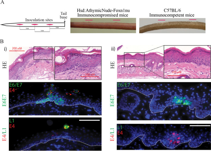
Identification of productive MmuPV1 lesions in immunocompetent mice. (A) Diagrammatic representation of inoculation site procedure in our model (left panel). Inoculation sites are shown in red, and the 1-cm spaces between the center of each wound site are annotated. The first wound site is located 1 cm down from the base of the tail. There is early visible lesion formation on immunodeficient nude mouse tail 10 days postinfection: sites are indicated with red lines (center panel). C57BL/6 immunocompetent mice showed no evidence of papilloma formation at wound sites 10 days postinfection (right panel). (B) Transient lesions located in C57BL/6 immunocompetent mouse tail tissue wound site 7 and 10 days following inoculation (i and ii). Panels show HE staining (top panel), E6/E7 RNAScope immunofluorescence and E4 protein (center), and immunofluorescent detection of E4 and L1 proteins (bottom panel). The nuclei were counterstained with DAPI. The scale is shown with a white bar (100 μm). The boxed areas in HE staining are enlarged. The dotted lines indicate the position of the basal layer.

