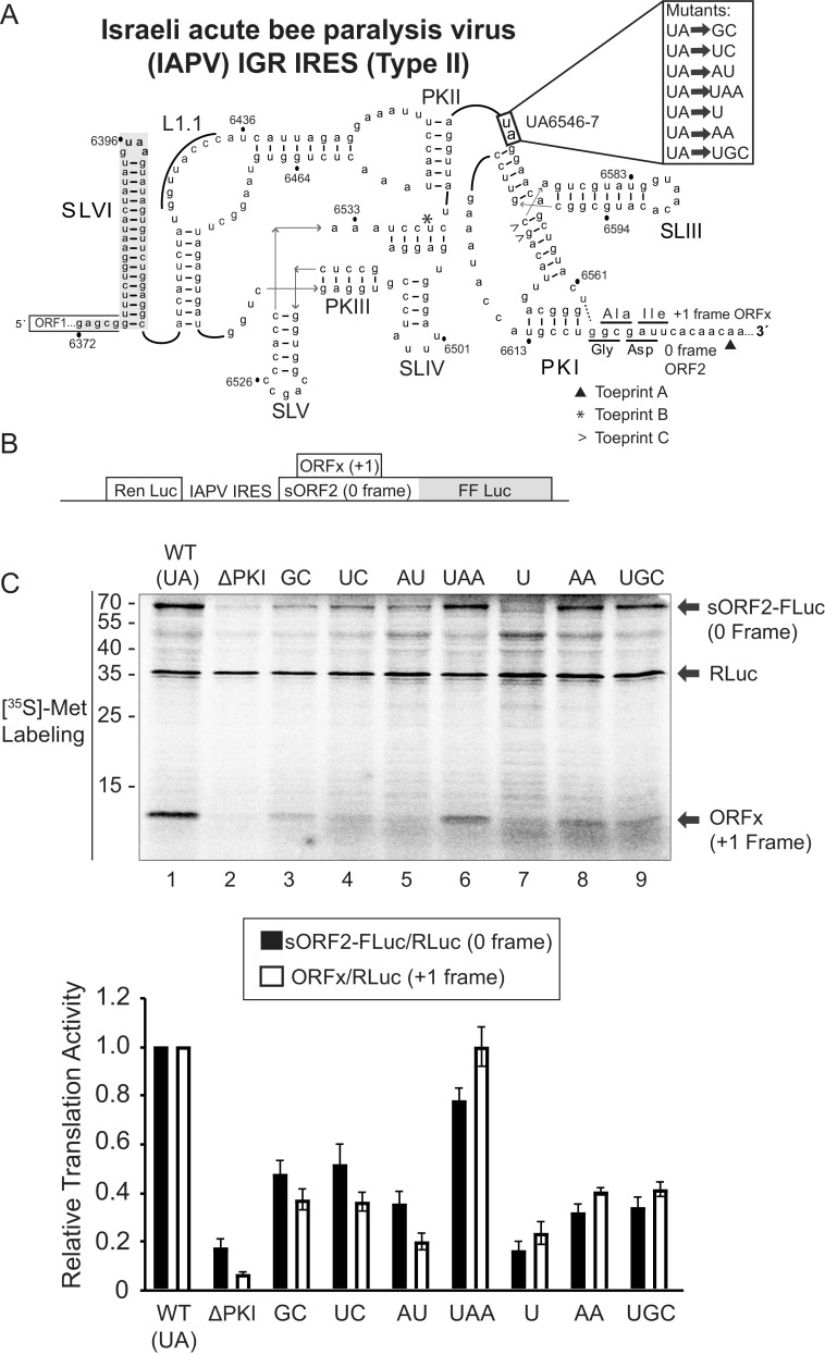
FIG 1.
Mutational analysis of the hinge region of the IAPV IGR IRES. (A) Schematic of the secondary structure of the IRES within the intergenic region of the Israeli acute paralysis virus genome. Key stem-loops (SL) IV, V, and VI and pseudoknots (PK) I, II, and III are shown. The hinge mutations are highlighted to the right. U6562-G6618 wobble base pair (dashed line) mediates +1 frame translation ORFx. (B) Schematic of the bicistronic IRES reporter construct. The renilla luciferase reporter (RLuc) and firefly luciferase reporter (FLuc) are translated via scanning-dependent and IRES-dependent translation, respectively. Fluc is fused in the 0-reading frame in-frame with a short ORF2 sequence (sORF2) (71.1 kDa). Expression of the +1 frame ORFx produces an ∼11.1 kDa protein. (C) Translational activities of mutant IRESs. (Top) Bicistronic reporter constructs were linearized and incubated in Sf21 extracts for 120 min at 30°C in the presence of [35S]-methionine/cysteine. Reactions were analyzed by SDS-PAGE, followed by phosphorimager analysis. A representative gel is shown. (Bottom) Quantitations of radiolabeled protein products. The ratios of sORF2-FLuc (0 frame)/RLuc and ORFx (+1 frame)/RLuc normalized to wild-type 0 and +1 frame translation are shown. Averages are from at least three independent experiments ± SD are shown.

