. 2020 Nov 13;12(3):983–992. doi: 10.1039/d0sc05064d

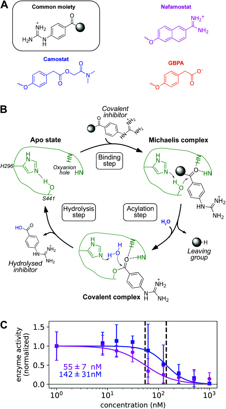
Fig. 2. (A) Chemical structure of nafamostat (magenta), camostat (blue), and GBPA (orange), split in a common moiety (4-guanidinobenzoyl) and different leaving groups. Note that GBPA is the hydrolyzed version of camostat's leaving group ester. (B) General mechanism of serine proteases applied to the hydrolysis of 4-guanidinobenzoyl esters by TMPRSS2. Only H296 and S441 residues of the catalytic triad and the two backbone NH groups of the oxyanion hole are depicted for clarity (enzyme color coded in green). (C) Dose response behavior of TMPRSS2 inhibition by nafamostat (magenta) and camostat (blue) with IC50s (data normalized, background subtracted). Experimental enzyme activities are reported at different drug concentrations as mean and standard deviation across independent experiments, continuous lines depict fitted dose–response model used for IC50 computation.

Fig. 2