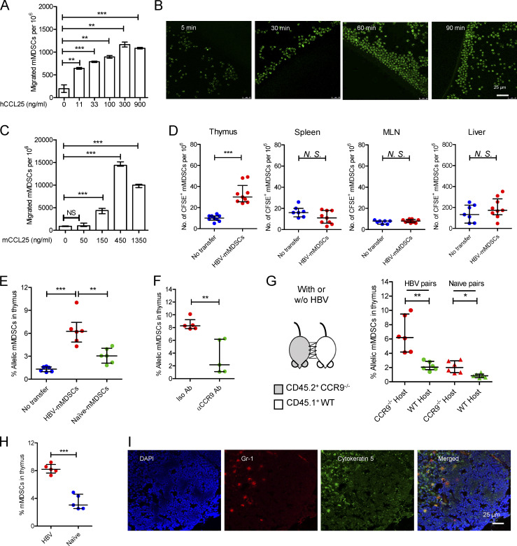
Figure 3.
Thymic homing of mMDSCs. (A) Transwell migration study of mMDSCs from CHB patients toward hCCL25 (n = 3). (B) Time-course migration study of mMDSCs toward agarose dots containing hCCL25, triplicate. (C) Transwell migration study of PBLs from HBV-persistent HDI mice (n = 3). (D) Separately detecting mMDSCs in different tissues from naive mice at 36 h after ADT with CFSE-labeled mMDSCs from HBV-persistent HDI mice (107 cells per mice, n = 9). (E) Detecting allelic cells via flow cytometry after ADT of CD45.2+ HBV-persistent HDI– or naive mice–derived mMDSCs to CD45.1+ mice (n = 6). (F) Detecting mMDSCs in thymi of CD45.1+ mice after transfer of CCR9 antibody–blocked CD45.2+ mMDSCs (n = 5). (G) Detecting thymic allelic mMDSCs in pairs of WT and CCR9 knockout (CCR9−/−) HBV or naive mouse pairs from parabiosis study (n = 6). (H) Detecting endogenous mMDSCs in the thymi from HDI-persistent HBV C57BL/6 mice (n = 5). (I) Location of endogenous mMDSCs in the thymi of HBV-persistent HDI mice. Nucleus (DAPI, blue), mMDSCs (Gr1, red), thymic medulla (cytokeratin-5, green), IF, triplicate. *, P < 0.05; **, P < 0.01; ***, P < 0.001.

