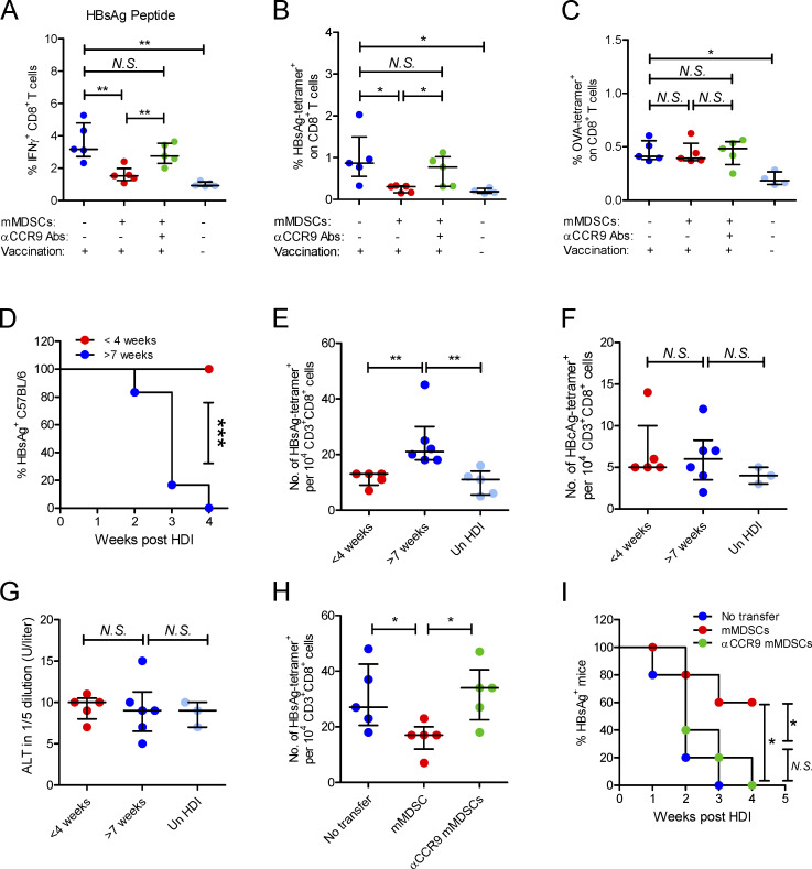
Figure 6.
mMDSCs mediate HBsAg-specific CD8+ T cell deletion and HBsAg persistence through CCR9. (A) Intracellular staining IFN-γ–positive CD8+ T cells in HBsAg peptide-stimulated (5 μg/ml) PBLs from HBsAg-vaccinated mice after transfer of HBV-persistent HDI mice–derived mMDSCs (n = 5). (B) Detecting HBsAg-specific CD8+ T cells with the MHC tetramer in splenocytes of HBsAg-vaccinated mice (n = 5). (C) Detecting OVA-specific CD8+ T cells in splenocytes of OVA-vaccinated mice after transfer of HBV-persistent HDI mice–derived mMDSCs (n = 5). (D) Persistence of serum HBsAg in C57BL/6 mice with different ages (n = 6). (E–G) The levels of HBsAg-specific CD8+ T cells (E), HBcAg-specific CD8+ T cells (F), and ALT (G) in C57BL/6 mice with different ages, day 10 after HDI of HBV B6 strain (n = 5). (H) Level of HBsAg-specific CD8+ T cells in mice after pretransfer of HBV B6 HDI mice–derived mMDSCs (n = 5). (I) Persistence of serum HBsAg in mice with pretransfer of mMDSCs (n = 5). *, P < 0.05; **, P < 0.01.

