
Figure 2.
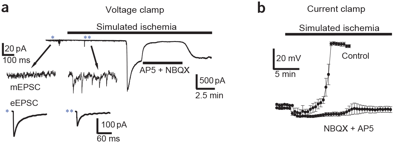
Simulated ischemia affects three aspects of glutamatergic signaling. (a) Voltage-clamp recording (holding voltage, Vh = −30 mV) of current in a CA1 pyramidal cell in a hippocampal slice during simulated ischemia. Blockade of large inward current by NMDA receptor antagonist AP5 (50 μM) and non-NMDA receptor antagonist NBQX (25 μM) indicates that it is generated by glutamate receptors. Inset shows typical examples of changes (*before ischemia compared with **after ischemia) in miniature EPSC (mEPSC) frequency (top) and suppression of evoked EPSC (eEPSC) amplitude (bottom) from different cells. Traces are adapted from data gathered in ref. 9. (b) Plot shows average (n = 3 or 4) membrane potential of Purkinje cells in cerebellar slices during simulated ischemia under control conditions or in the presence of glutamate receptor antagonists (as in a). Data from ref. 69.