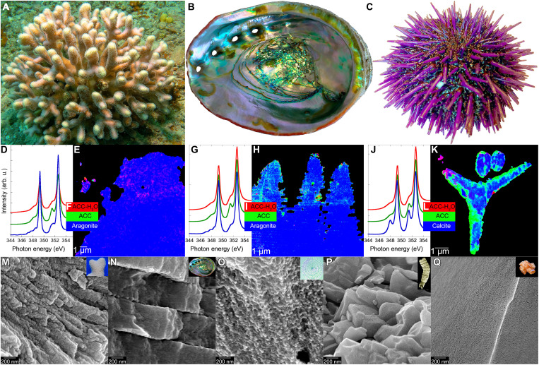
Fig. 2. The same amorphous precursors across phyla: Cnidarians, mollusks, and echinoderms.
(A) S. pistillata coral in the Red Sea (photo credit: T.M.). (B) California red abalone Haliotis rufescens (photo credit: P.U.P.A.G.). (C) California purple sea urchin Strongylocentrotus purpuratus (photo credit: P.U.P.A.G.). (D, G, and J) X-ray absorption spectra from nanoscale regions of fresh forming biominerals: S. pistillata skeleton, H. rufescens nacre, and S. purpuratus embryonic spicules. Three distinct spectral line shapes at the Ca L-edge, and thus, three distinct mineral phases or “components” occur in each biomineral: hydrated ACC, anhydrous ACC, and crystalline calcite or aragonite. (E, H, and K) Component maps showing abundant amorphous pixels in the forming parts of each biomineral and submicrometer amorphous particles in nearby cells. (F, I, and L) Color legend for both component spectra (D, G, and J) and component maps (E, H, and K). (M to P) Scanning electron micrographs showing that modern and fossil biominerals show nanoparticulate texture after cryofracturing (M to P), whereas nonbiogenic minerals do not (Q). Insets in (M) to (Q) show photographs of each sample. (M and N) Modern aragonite biominerals: coral skeleton from S. pistillata (M) and nacre from H. rufescens (N). (O) Calcite sea urchin spine from S. purpuratus. (P) Phosphatized Ediacaran Cloudina (550 Ma before present) from Lijiagou, China. (Q) Nonbiogenic aragonite from Sefrou, Morocco. Data are from (15, 23–25). arb. u., arbitrary units.

