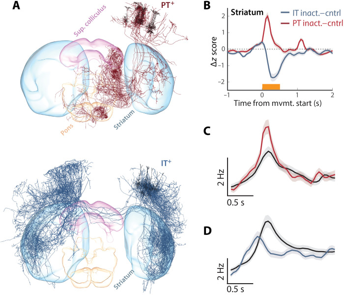
Fig. 8. Inactivation of PT and IT neurons oppositely affect striatal activity.
(A) 3D visualization of complete single-neuron reconstructions (3) for 10 representative single-cell reconstructions of layer 5 PT (top) and IT (bottom) anatomical classes from the mouselight.janelia.org database shows partially overlapping projections to the recorded region of dSTR. (B) For all units from dSTR, we computed the difference between movement-aligned, z-scored time histogram for control trials and perturbation trials in which either Sim1+ corticopontine PT neurons (red) or layer 5 Tlx3+ IT neurons (blue) were inactivated during movement. (C and D) Populations of units with low average firing rates (also broad spike widths on average) were used to assess whether modulation of dSTR activity was consistent with changes in medium spiny projection neuron activity during PT (C) or IT (D) inactivation trials. Control trials (black) reveal clear movement-aligned modulation of activity in these populations and opposing changes during inactivation.

