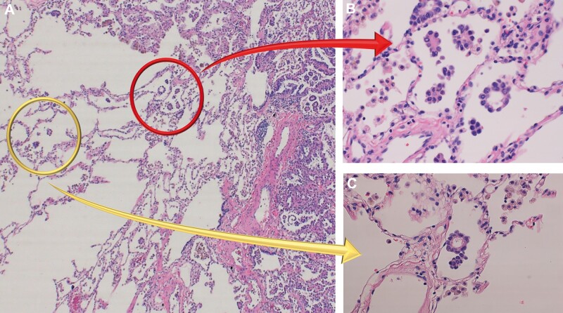
Figure 1:
Pathological findings of spread through air spaces (STAS). (A) High-power field image (×100) of lung adenocarcinoma. Tumour margin and normal alveolar spaces are observed. (B) STAS adjacent to the edge of tumours (red arrow). STAS are defined as (i) micropapillary structures consisting without central fibrovascular cores, (ii) solid nests or tumour island filing air spaces, and (iii) single cells consisting of scattered discohesive single cells. (C) STAS found within air spaces in the lung parenchyma beyond the edge of the main tumour (yellow arrow).

