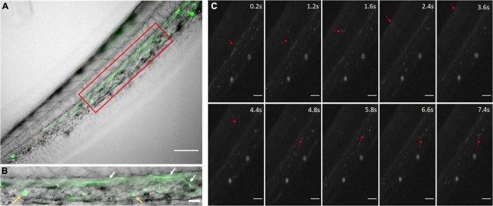
FIGURE 2.
Coumarin-6 Loaded Nanoparticles after Injection. (A) Coumarin-6 nanoparticles traveling in the caudal artery of the zebrafish tail. Red box is blown up and shown as (B). White arrows show nanoparticles rolling along vessel walls. Yellow arrows indicate clusters of endocytosed nanoparticles. (C) Representative stills showing nanoparticles traveling from the caudal artery (0.2–1.2 s), into the capillaries of the tail tissue (1.6–4.8 s) and entering the posterior caudal vein (6.6–7.4 s). All scale bars represent 100 μm.

