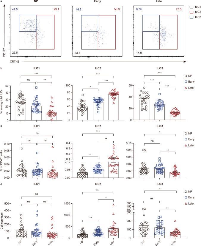
Fig. 2.
ILC subsets distribute dynamically in peripheral blood of non-pregnant women and pregnant women of different trimesters. a–d Analysis results for ILC subsets in peripheral blood samples of non-pregnant, early-pregnant, and late-pregnant women. a Representative flow cytometry plots are shown in which numbers indicate the frequency of flow cytometric events. Comparison of proportion of ILC subsets within total ILCs (b), ILC subset percentage in CD45+ cells (c) and absolute counts per milliliter of blood (d) in peripheral blood of non-pregnant women (black circles), early-pregnant women (blue squares), and late-pregnant women (red triangles). Data are shown as means ± SEMs and were analyzed by Kruskal-Wallis tests. Each point indicates an individual. *P < 0.05, **P < 0.01, and ***P < 0.001. ns, not significant

