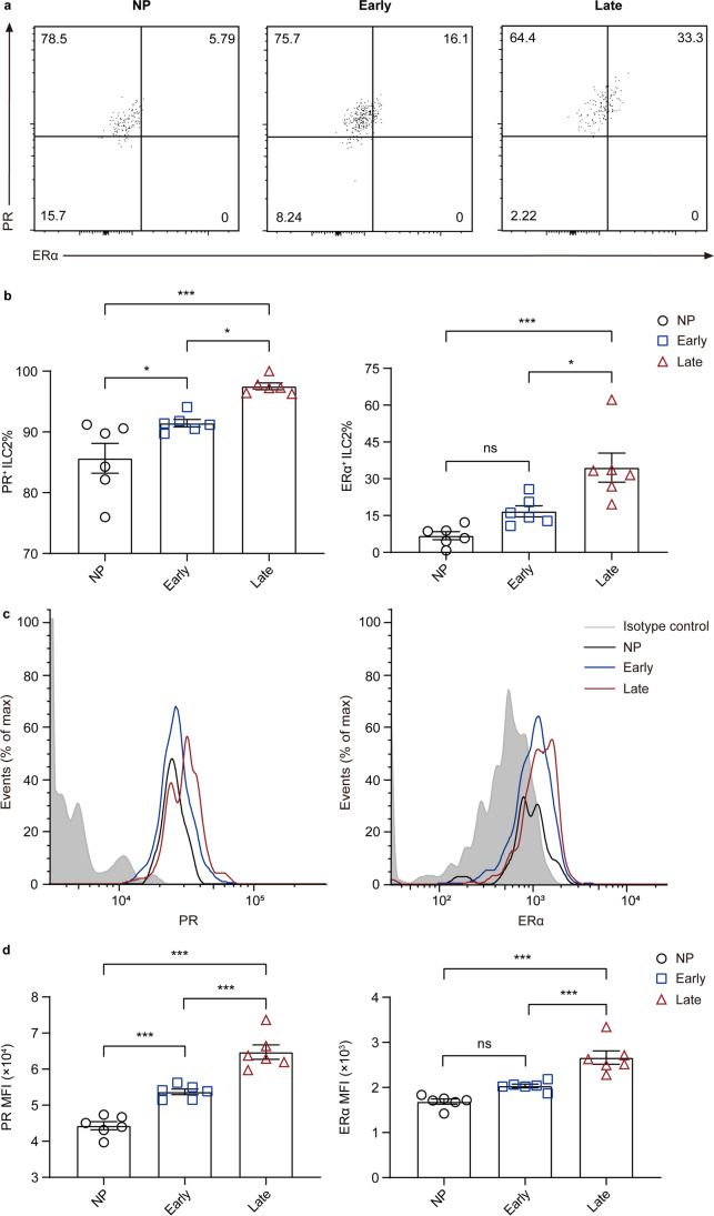
Fig. 4.
The estrogen and progesterone receptors in circulating ILC2s change differently in non-pregnant and pregnant women of different trimesters. a–d Flow cytometric analysis for PR and ERα in peripheral ILC2s of non-pregnant, early-pregnant, and late-pregnant women. a Representative flow cytometry plots are shown in which numbers indicate the frequency of flow cytometric events. b Comparison of and PR+ ILC2 and ERα+ ILC2 proportion in peripheral blood of non-pregnant women (black circles), early-pregnant women (blue squares), and late-pregnant women (red triangles). c Representative flow cytometry histograms of intracellular PR and ERα in circulating ILC2s from isotype control (grey area), non-pregnant women (black lines), early-pregnant women (blue lines), and late-pregnant women (red lines). d Quantification of PR and ERα mean fluorescence intensity (MFI) of circulating ILC2s. Data are shown as means ± SEMs and were analyzed by one-way ANOVA. *P < 0.05 and ***P < 0.001. ns, not significant

