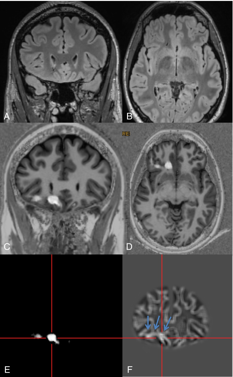
Fig. 2.
A 28-year-old man with hypomotor seizures and clonic seizures with versive head movement to the left side evolving to bilateral tonic–clonic seizures (#40). MRI with coronal (A) and axial reformations (B) of a 3D FLAIR sequence was considered to be normal. By scrolling through the co-registered MP2RAGE images (C, D), the lesion was detected “within a minute.” For co-registration, the ANN probability map (E) is used. The junction image as one of the input maps for the ANN highlights the blurring of the gray white matter junction as the most prevalent feature of FCD (F: arrows). Epileptogenicity of the lesion was confirmed by SEEG, the patient underwent surgery, and histopathology revealed a mild malformation of cortical development (mMCD)

