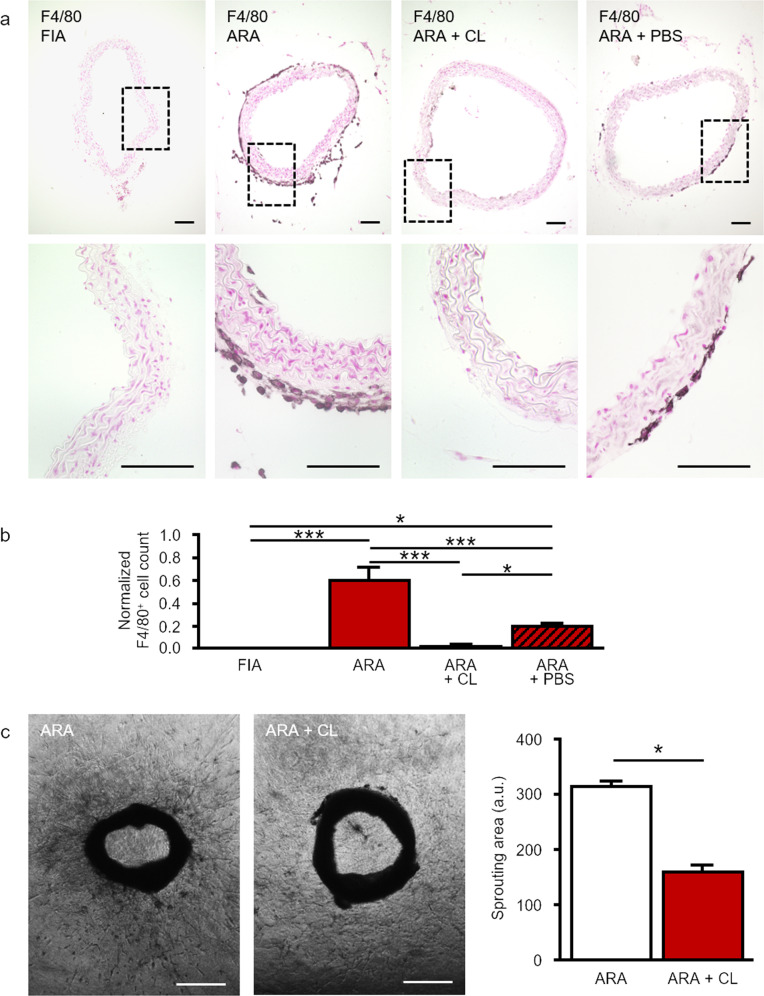
Fig. 1. Generation of F4/80+ macrophages in ARA.
a Cross-sections of murine aortae were immunostained for the marker of mature macrophages F4/80. In FIA, hardly any F4/80+ macrophage could be found. After ARA a few F4/80+ macrophages appeared within the intima. However, within the adventitia, the number of F4/80+ macrophages increased substantially. Compared to control, cultured aortic rings treated with clodronate-containing liposomes displayed almost no F4/80 immunostaining due to efficient depletion of F4/80+ macrophages within the adventitia. b Statistical analysis of the number of adventitial F4/80+ macrophages with and without liposome treatment. Application of PBS-containing liposomes had a less pronounced effect compared to clodronate-containing liposomes. Cell counts were normalized to total media area (n = 7–10). c Phase-contrast images and quantification of sprouting area of cultured aortic rings with or without clodronate treatment. Depletion of macrophages by clodronate-containing liposomes reduced total cellular sprouting as well as capillary-like tube formation (n = 23–31). *P < 0.05; ***P < 0.001. Scale bars: in a 100 µm, in c 500 µm. ARA aortic ring assay, ARA + CL ARA with clodronate-containing liposomes, ARA + PBS ARA with PBS-containing liposomes.

