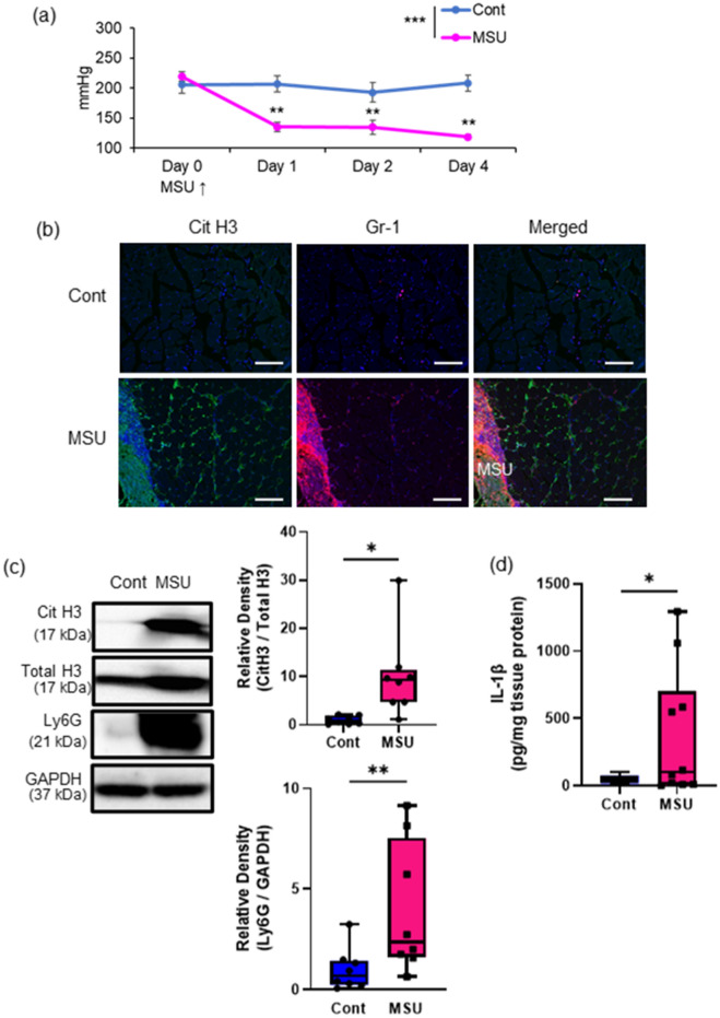
Figure 1.
Induction of muscle hyperalgesia and NETs with intramuscular MSU injection. (a) Time-series experiment of the MNT values in the MSU- or saline-injected (contralateral) hindlimbs (n = 6 in each group). (b) Representative IHC images (200 ×) of TSM tissue on day 2, indicating citH3 (green), Gr-1 (red), and DAPI (blue). (c) Western blotting analysis of citH3 and Ly6G amounts in the TSM tissues on day 2 after intramuscular injection of MSU or saline (n = 8 in each group). The relative density is normalized based on the loading control (total histone H3: total H3 and GAPDH, respectively). Scale bar = 100 μm. (d) ELISA analysis was performed to evaluate protein levels of IL-1β, using MSU- or saline-injected TSM tissues on day 2 (n = 10 in each group). All data are shown as Tukey’s boxplots with individual data points and analyzed using paired t-test or two-way ANOVA with Tukey’s post-hoc multiple comparison test for time-series of MNT values. Statistical significance is indicated with * (p < 0.05), ** (p < 0.01), and *** (p < 0.001).

