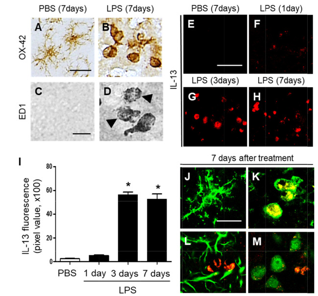
Fig. 2.
Interleukin-13 expression in the striatum of LPS-injected rat in vivo. (A~D) Striatal sections (A, C, PBS; B, D, LPS) adjacent to those used in Fig. 1A and C were immunostained with OX-42 (A, B) and ED1 (C, D) antibodies for microglia. Accumulating intracellular vacuoles are denoted by arrowheads in D. (E~H) IL-13 immunofluorescence staining in the striatum at 7 days after intrastriatal injection of PBS as a control (E), and at 1 day (F), 3 days (G), or 7 days (H) after intrastriatal injection of LPS. (I) Quantification of IL-13 expression in OX-42+ cells in the striatum at indicated time points. *p<0.001, as compared with PBS. One-way ANOVA and Newman–Keuls analyses. Seven animals were used for each experimental group. The results represent mean ± SEM. (J~M) Immunofluorescence images of interleukin 13 (IL-13, K; red) and OX-42 (J, K; green), or IL-13 (L; red) and GFAP (L; green), or IL-13 (M; red) and NeuN (M; green) and both images are merged (Yellow; K) in the striatum at 7 days after PBS (J) or LPS (K~M) injection. Scale bars, (A, B) 25 µm; (C, D) 20 µm; (E~H) 50 µm; (J~M) 20 µm.

