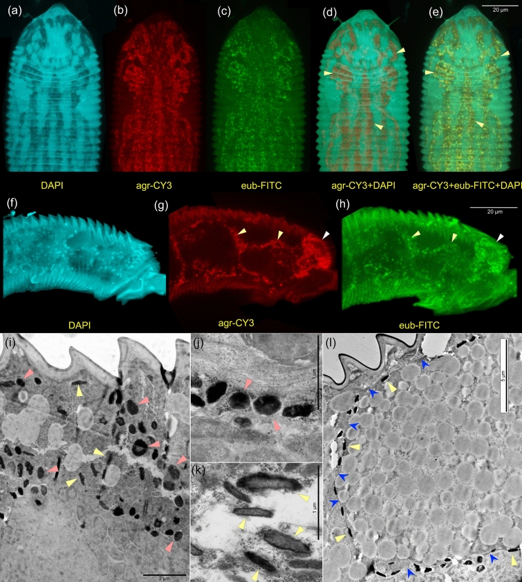
Figure 4.
Endosymbiotic bacteria of the mite Fragariocoptes setiger, fluorescence in situ hybridization (FISH) with different fluorophores and oligonucleotide probes (a–h) and TEM microscopy (i–l). Mite anterodorsal (a–e) and anterolateral parts (f–h); intermuscular bacteria (d-e, yellow arrowheads), bacteria surrounding gigantic parenchymal cells (g, h, yellow arrowheads) and salivary glands (g, h, white arrowheads); DAPI + no probe (a, f); CY3 + 16S.1722F.Agr.tum (b, g); FITC + Eub338 (c, g); CY3 + 16S.1722F.Agr.tum, DAPI (d); CY3 + 16S.1722F.Agr.tum, FITC + eub338, DAPI (e). Bacterial morphotype 1 (Wolbachia) (i, j, red arrowheads), and bacterial morphotype 2 (yellow arrowheads) (k, l, yellow arrowheads) in various locations inside the mite: mid-lateral opisthosoma with saw-like cuticle and underlying tissues are visible (i), same as previous, a gigantic parenchymal cell (fat body) is shown and traced by blue arrows (l), spaces between the fat body and the gut (j, k).

